*Corresponding Author:
Ana Margarida Campos Cerqueira,
Department of Internal Medicine, Hospital Senhora da Oliveira, Guimaraes, Portugal
Tel: +351 253540330
Email: migui_campcer@hotmail.com
Abstract
Here in we present you a clinical image of a metastatic liver. Our patient was a 58 year old male, with past smoking habits, that went to our ER complaining about vomiting and asthenia within the last week. While oh the Internal Medicine infirmary he did an ultrasound guided biopsy that confirmed pulmonary origin of the cancer. He was transferred to the Pneumology infirmary and continued the staging of his disease. At the end, were also documented brain metastases and he was referred for palliative care.
Clinical Image
We report a case of a 58-year-old Caucasian male with a past medical history of chronic obstructive pulmonary disease, cardiac insufficiency, 2 strokes without sequelae, and past smoking habits. He was admitted at out ER for vomiting and asthenia within the last 7 days. An abdominal CT scan was performed at admission with documentation of 3 large hypodense and hypocaptant lesions on his liver, the largest being in the right lobe (10 cm), which suggested a secondary origin. He was admitted at our Internal Medicine ward to run more studies to clarify the cause of these hepatic lesions. Ultrasound guided biopsy was performed with histological confirmation of primary Lung Cancer and the patient was transferred to Pneumology infirmary. Brain metastases have also been documented. He was oriented towards Palliative care (Figure 1)
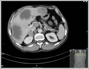
Figure 1: Mtx-Hepáticas
Citation: Cerqueira AMC, de Melo Seco T, Cotter J (2019) When the Liver Tells you to Look at the Lungs. J Case Repo Imag 4: 011.
Copyright: © 2020 Cerqueir AMC, et al. This is an open-access article distributed under the terms of the Creative Commons Attribution License, which permits unrestricted use, distribution, and reproduction in any medium, provided the original author and source are credited.
