*Corresponding Author:
SZ Mavlyanova,
Republican specialized scientific-practical medical center of dermatovenereology and cosmetology of the Ministry of Health of the Republic of Uzbekistan
Tel: +998901885779
E-mail: shahnoza_m@mail.ru
Abstract
The article presents a clinical and immunological study of auto- immune antibodies of the IgG class to denatured and undenatured DNA and C-reactive protein in patients with acantholytic pemphigus. The results of the study showed. that in patients with serum with AP there is a development of an autoimmune process of a systemic nature, characterized by a high concentration of autoantibodies (AAT) of IgG class to ds-DNA by 1.9 times and C - reactive protein by 2.02 times compared to control healthy individuals.
Keywords
Pemphigus, Antibodies, C-reactive protein
Introduction
In recent times was fixed in increase in number of patients with acantholytic pemphigus (AP) among severe forms of skin diseases. Pemphigus is one of severe autoimmune skin diseases and the mucous membranes in which formed autoantibodies of immunoglobulin of IgG class to desmogleins 1 and 3. The given proteins belong to super-family of calcium-dependent molecules of cellulat adhesion that are involved in intercellular interactions [1-5]. Herewith a level of acantholysis in different forms of autoimmune pemphigus explains by location of desmogleins 1 and 3 in skin epidermis and epithelium of mucous membrane of oral cavity [1].
Studying mechanisms of development and treatment of this extremely seriosus skin disease by its actuality takes one of the leading places in dermatology practice. In pathogenesis of acantholytic pemphigus an important place allocated autoimmune mechanisms. Literature data exhibited that in mechanism of development of autoimmune antibodies a special role may play antibodies to double-stranded DNA (ds-DNA) thatis responsible for autoimmune process in organism. It should be noted that antibodies to double-stranded DNA (ds-DNA) belong to a group 0f antinuclear antibodies that is autoantibodies directed by organism against components of its own nuclei. Frequency of occurrence of these AT is well above in patients with an unfavorable course of disease [2], as well as it takes place in dynamic observation of active phase of disease [1] that indicates a close relation of production of АТ with main stages of development of disease.
Levels of antinuclear antibodies may vary considerably not only in dependence on individual immune reactivity of patient, on character and stage of disease but also as fact of autoimmune answer haw native and single-stranded DNA [1].
It is shown that antibodies to native and one-stranded DNA may play a role in development of such autoimmune diseases as systemic lupus erythematosus (SLE), disseminated sclerosis, autoimmune thyroiditis. Moreover, detection of ds-DNA is one of criteria to diagnose SLE [6-9].
С-reactive protein (C-RP) is multi-functional protein of acutephase, playing an important role in protection from alien agent in autoimmune processes. It is included in non-specific answer immediately after penetration of antigen in organism and acts through stimulation of phagocytosis of neutrophils and cells of macrophages range [10,11].
Material and Methods
Our studies were aimed at Estimation of importance of the revealed autoantigens of IgG class to double-stranded DNA (ds-DNA), autoantigens of IgG class to single-stranded DNA (ss-DNA) and SRB in clinical course of acantholyticpemphigus.
Eighty-six patients with pemphigus aged from 18 to 71 years old of IgG class to single-stranded (ss-DNA) double-stranded (ds-DNA) and concentration of C-reactive protein (CRP) in blood serum were determined by means of enzyme-linked immunosorbent assay (ELISA) ( of Vector-Best», Russia). All the patients were consulted by related specialists: therapist, endocrinologist and other.
Result
Result of 86 pemphigus patients pemphigus vulgaris (P.vulgaris) was diagnosed in 80 (91.9%), p. erythymatosus, seborrthoicum – in 5 (5.7%) and p.vegetans– in 1 (2.3%) respectively. In all the patients in lesion foci in content of bubbles/or erosive areas cytology examination revealed cells of acantholytic cells.
Results of study showed reliable increase of autontibodies (AAB) of IgG class to double-stranded DNA (ds-DNA). Of 86 patients in 48 patients was noted an increase in level of AAB IgG to ds-DNA that formed 55.8% cases. Whereas AAB IgG to ss-DNA were revealed 22 patients that formed 25.6% cases. In group of healthy persons among 18 only one had an increased level of AAB IgG to ss-DNA that formed 5.8% cases (Figure1). Results obtained evidenced development оf autoimmune process in patients of acantholytic pemphigus.
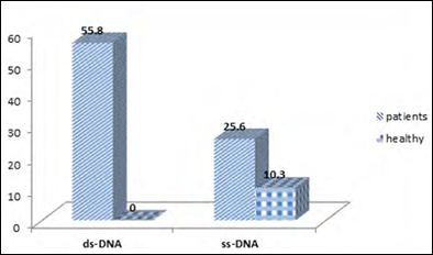
Figure 1: Index of frequency of the detection of ds-DNA and ss-DNAin blood serum of patients with pemphigus (abs, %).
Studying concentration of AAB of IgG class to ds-DNA in blood serum of patients with pemphigus exhibited a reliable increase of indications by 1.9 times as compared with control group that formed on average 29.9 ± 0.8 Еd/ml. While concentration of ss-DNA on average compiled 27.01±1.4 Ed/ml that by 1.5 times exceeded performance of healthy persons. Data obtained had statistically reliable character (Р <0.05).
As a rule, a high index of AAB IgG to double(ds-DNA) and single-stranded DNA (ss-DNA) in blood serum evidenced an increased activity of acantholytic pemphigus, acceleration of inflammatory process in organism of patients with pemphigus.
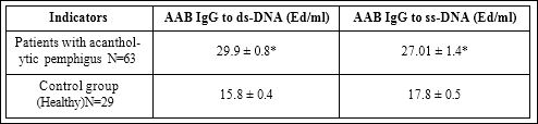
Table 1: Indicators of AAB IgG to denatured DNA (ss-DNA) and non-denatured DNA (ds-DNA) in patients with pemphigus (М+m).
Note: *Index of reliability towards indicators of healthy persons (Р<0,05).
State of concentration of ААB IgG to ds-DNA and ААB IgG to ss- DNA in blood serum in dependence on duration of the disease (Table 2) was analyzed by us.
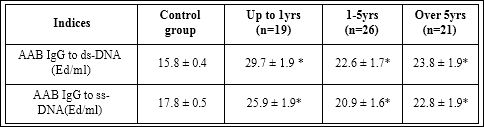
Table 2: State of concentration of ААB IgG to ds-DNA and ААB IgG to ss-DNA in blood serum of patients with pemphigus given the duration of the disease (М+m).
Note: *Index of reliability towards indicators of healthy persons (Р <0.05).
As follows from Table 2, concentration of ААB IgG to ds-DNA in patients with AP up to 1 year averaged 29.7 ± 1.9 МЕ/ml, in duration 1-5 years- 22.6 ± 1.7 Еd/ml and in duration over 5 years-23.8 ± 1.9 Еd/ ml respectively. Whereas concentration of AAB IgG to ss-DNA up to 1 year averaged 25.9 ± 1.9 Еd/ml, and with increasing prescription averaged – 20.9 ± 1.6 and 22.8 ± 1.9 Еd/ml respectively.
Data obtained evidenced that initial period of morbidity was characterized by expression of autoimmune process that causing by a high concentration of AAB IgG to ds-DNA by 1.3 times compared with indices od duration of disease 1-5 years and over 5 years respectively. Similar indictors were observed in AAB IgG to ss-DNA. Decline in performance of concentration ААB IgG to ds-DNA and ААB IgG to ss-DNA with increase of duration of disease, in our opinion, is related with taking glucocorticosteroids therapy. However, it should be noted, that concentration of AAB against a background of hormonal therapy remained relatively high compared with indicators of control healthy group (Р<0.05). Data retrieved evidenced preservation of activation of autoimmune process, development opportunity of complicated somatic and opportunistic infection processes in organism of patients with AP.
Study of C-reactive protein (C-RP) in blood serum of patients with AP, relating a group of proteins in acute phase, revealed an increase of concentration by 2.02 times that on the average was14.4 ± 0.7 МЕ/l compared with control group (Table 3).
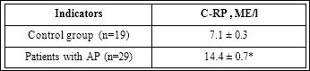
Table 3: Indicators of CRP in patients with acantholytic pemphigus.
Note: *Index of reliability towards indicators of healthy persons (Р <0.05).
As follows from table 3, an increased concentration of C-reactive protein in blood serum evidenced an expression of inflammatory process in organism of patients with AP. Analysis of results taking into consideration duration of disease revealed an increased concentration of C-reactive protein in blood serum of patients that in our opinion, is associated with autoimmune process. At the same time with increase of duration of disease a level of C-reactive protein remained on enough high level and averaged 15.8 ± 1.8 МЕ/l (Р<0,05) (Table 4).
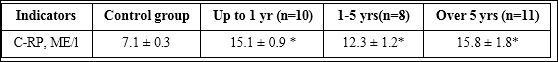
Table 4: State of C-RP in blood serum of patients with AP taking into account duration of disease (М+m)
Note: *Index of reliability towards indicators of healthy persons(Р <0.05).
Therefore, reliable increase of AAB concentration of IgG class to non-denatured DNA (ds-DNA) and to denatured DNA (ss-DNA) noted to be in patients with acantholytic pemphigus in blood serum 1.9 and 1.5 times as compared with control group that reflects expression of autoimmune process on organism of patients. The level of C-reactive protein in blood serum of patients with AP belonged to group of proteins of acute phase averaged 14.4± 0.7 МЕ/l that is by 2.02 times higher that indicators of control group. Data received evidenced expression of inflammatoru process against a background of immune process.
Conclusion
- Development of autoimmune process of systemic character was noted in patients with AP characterized by a high detection of an- tibodies (AAB) IgG class to double-stranded DNA (ds-DNA) in 55.8% cases.
- Taking into account duration of disease expression of autoimmune process most frequent manifested up to 1 year characterized by increase of IgG concentration to non-denatured DNA (ds-DNA) 1.8 times as compared with indicators of control healthy persons.
References
- Lysenko АА (2009) Analysis of a role of human distal domains of desmoglein 3 in disturbance adhesion between keratinocytes in pemphigus.
- Makarova YaYu (2005) Estimation of role of immune status in development of complications of corticosteroid therapy of authentic acantholytic pemphigus
- Reshetnikova (2005) Т.B Role of immunopathologic processes in genesis of authentic acantholytic pemphigus and methods to correct them.Thesis 34.
- Тeplyuk NP, Lepechova АA (2014) Clinical aspects of steroid resistance in autoimmune Russian J of Skin & Venereal Dis2:3-6.
- Christen U, von Herrath MG (2004) Initiation of autoimmunity. Curr OpinImmunol 16: 759-767.
- Gill JM, Quisel AM, Rocca PV, Walters DT (2003) Diagnosis of systemic lupus Am Fam Physician 68: 2179-2186.
- Fauci AS, Kasper DL, Longo DL, Braunwald E, Hauser SL, et al. (2008) Harrison’s Principles of Internal Medicine. 17 ed. – The McGraw-Hill Companies.
- Nossent HC, Rekvig OP (2005) Is closer linkage between systemic lupus erythematosus and anti-double-stranded DNA antibodiesa desirable and attainable goal? Arthritis Res Ther 7: 85-87.
- Russell AI, Cunninghame Graham DS, Shepherd C, Roberton CA, Whittaker J, et (2004) Polymorphism at the C-reactive protein locus influences gene expression and predisposes to systemic lupus erythematosus 13: 137-147.
- Bongu A, Chang E, Ramsey GR (2002) Can morbidity and mortality of SLE be improved?. Best Pract Res Clin Rheumatol 16: 313-332.
- Pepys MB, Hirschfield GM, Tennent GA, Gallimore JR, Kahan MC (2006) Targeting C-reactive protein for the treatment of cardiovascular disease. Nature 440: 1217-1221.
Citation: Mavlyanova SZ, Alimukhamedova YA, Abdukadirov AO, Kodirova MA (2019) To the state of the autoimmune process in patients with acantholytic pemphigus. J Cell Mol Biol 3: 008.
Copyright: © 2019 Mavlyanova SZ, et al. This is an open-access article distributed under the terms of the Creative Commons Attribution License, which permits unrestricted use, distribution, and reproduction in any medium, provided the original author and source are credited.