*Corresponding Author:
Andrey N Belousov,
Laboratory of Applied Nanotechnology of Belousov, Kharkov Medical Academy, pr. Nauky, 31-v, fl 32, Kharkov, 61072, Ukraine
Tel: +380 509151889
E-mail: an.belousov2012@yandex.ua
Abstract
The main purpose of this work is inhibition of erythrocytes hemolysison the whole blood by means of magnetite nanoparticles (magnet-controlled sorbent MCS-B). Conventional erythrocytes of person’s venous blood were objects of the research. The time of appearing the signs of erythrocytes hemolysis was recorded with the help of visual method. In result of investigation it was established that magnetite nanoparticles not only reliably reduce hemolysis but also prolong time preservation of the blood, change activity adenosinetri phosphatases of erythrocytes and influence on transmembrane exchange, functioning of ion channels.
Keywords
Adenosinetriphosphatases; Haemolysis; Eryptosis; Nanoparticles (MCS-B); Transmembrane exchange
Introduction
Metabolic restoration, prolongation of normal function of cells both inside and outside the organism is the main purpose of medical and biological trend in the 21st century. With rapid progress in nanotechnology, many nano-size materials have been extensively used in biomedical and pharmaceutical industry and industrial production. Nevertheless, the rapid growth of nanotechnology has raised biological safety concerns because of the unique dimensional and physicochemical properties of the nano-size materials [1]. Although the mechanism of toxicity of nanomaterials is complicated and markedly different from that of traditional biomaterials, current evaluations of nanotoxicology are still confined to testing the compatibility of materials using traditional methodologies. A standard research protocol to evaluate the nanotoxicity is lacking, severely limiting the development and applications of nanoparticles [2].
Fe3O4 Magnetic Nanoparticles (Fe3O4-MNPs) is the only nanomaterial that has been approved for clinical applications because of their relative safety, unique magnetic responsiveness, and their simple and controllable preparation [3,4]. Although studies concerning the potential risks of Fe3O4-MNP have been reported the biocompatibility evaluation relies mainly on in vitro cytotoxicity such as hemolysis testing, cell viability, oxidative damage, inflammatory reactions and genotoxicity, or on pharmacokinetics, and in vivo bio-distribution [5,6].
Erythrocytes are the main components in the circulation system and are also one of the first components that Fe3O4-MNPs contact when the nanoparticles are administered through intravenous injection. Fe3O4-MNPs are generally regarded as hemocompatible based on very low hemolytic activity [7]. Hemolysis testing is a well-accepted classical assay for acute toxicity screening in evaluating hemocompatibility and can reflect the breakage to the erythrocyte membrane.
Accumulated evidence in recent years suggests that some nanoparticles have an adverse effect on erythrocytes pre-hemolysis after interacting with RBC in vitro and in vivo. Owing to the induction of specific structural changes in the lipid bilayer, these nanoparticles caused erythrocyte shape transformation decreased the deformability and oxygen-delivering ability modified the heme conformation and changed hemorheological properties [8-11]. Although it has been reported that MgNPs-Fe3O4 (100 mg/ml) can cause cellular membrane damage in cultured lung epithelial cells the impact of Fe3O4-MNPs at safe dose on cellular membrane of erythrocytes and circulatory properties beside hemolysis remains unknown [12].
The toxic effects determined based on the hemolysis, membrane injury, lipid peroxidation and antioxidant enzyme production were fairly size and dose dependent. For example, in particular, the smallest sized silver nanoparticles size of 15 nm (AgNPs15) displayed a greater ability to induce hemolysis and membrane damage than AgNPs50 and AgNPs100. Such cytotoxicity induced by AgNPs should be attributed to the direct interaction of the nanoparticle with the red blood cells (RBCs), resulting in the production of oxidative stress, membrane injury and subsequently hemolysis. Overall, the results suggest that particle size is a critical factor influencing the interaction between AgNPs and the RBCs [13].
In Ukraine, the first medical nanotechnology drug was synthesized and patented in 1998. These are such drugs as Intracorporeal Nanobiocorrector (ICNB), Magnet-Controlled Sorbent (MCS-B) and Micromage-B [14-16]. Basis of the drugs is magnetite of nanoparticles (Fe3O4) with the size ranging from 6 till 12 nm. Presence of adsorption layer provides high sorption activity for the magnetite of Nanoparticles (NPs). The total sorption surface of the magnetite NPs ranges from 800 to 1200 m2/g, and intensity of the magnetic field that induced by each magnetite NPs is 300-400 kA/m.
The main purpose of study was reduced hemolysis of erythrocytes on the whole blood by means of nanoparticles of Magnet-Controlled Sorbent (МСS-B).
To fulfill the aim the following tasks are to be solved:
- Determine the dependence between time of appearing hemolysis and amount of processing of blood with MCS-B
- Investigate activity of transport adenosinetriphosphatase of erythrocytes: Na, K АТPHase and Ca, Mg – АТPHase
- Investigate level of cytosolic calcium in erythrocytes
- Find optimum amount processing of blood by NPs of Magnet-Controlled Sorbent (MCS-B)
Material and Methods of Research
In order to determine ultrastructural reconstructions in hepatic cells under effect of magnetite, an experiment was conducted on 45 rabbits. In ear vein of the rabbit, from a calculation 6-8 ml/kg, 0.0225% was injected ICNB for 24 hours before the investigation. Physical and chemical properties of ICNB (Figures 1 & 2; Tables 1 & 2):
- Material: NPs of MCS-B. The basis of MCS-B is Fe3O4. Physical and chemical properties of MCS-B (Figures 1 & 2; Tables 1 & 2):
- 0.1% colloidal solution of magnetite nanoparticles
- Size of magnetite of nanoparticles is 6-12 nm (the data were quan- tified from TEM micrographs)
- Total area of surface magnetite of nanoparticles Ss = 800-1200 m2/g
- Magnetized of saturation is = 2.15 кА/m
- ζ Potential = - 19 mV
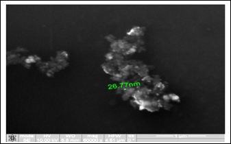
Figure 1: Study of magnetite nanoparticles (ICNB) with use microscope ion-electronic raster-type Quanta 200 3 D.
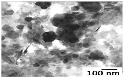
Figure 2: Study of magnetite Nanoparticles (ICNB) with use microscope electronic translucent JEM-2100.
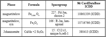
Table 1: X-ray analysis of ICNB in X-ray diffractometer RigakuUltima IV (CuKα, Kβ filter - Ni), one-coordinate DTeX semiconductor detector.
Table 2: The phases of ICNB (RIR - method; error 8± 3%).
Object of research: conventional erythrocytes venous on the whole blood of the person. All researches in vitro were performed. The condition of erythrocytes venous of blood in 20 healthy volunteers was studied. The age of persons varied from 24 to 40 years. The researches included 2 stages: stage I was condition of erythrocytes on the 1st day of observation; stage II was condition of erythrocytes on the 21th day of observation. The biochemical investigations were performed only on stage I. The estimate of visually state of erythrocytes (hemolysis) was performed on stages I and II.
Research methods: 12 ml venous of blood was taken from patient. For preventing coagulation of blood citrate sodium was introduced. Then in each tube 3 ml of blood was introduced. The first tube was control. In second tube of test was added MCS-B in quantity of 1.5ml with its following separation in during 30-40 sec by means of a constant magnetic field with the intensity 200 kA/m. In third tube of test the blood was twice processed by means of MCS-B. In fourth tube of test the blood was thrice processed by means of MCS-B. On stage I the activity of transport adenosinetriphosphatase (Na, K - АТPHase and Ca, Mg - АТPHase) and level of cytosolic calcium in erythrocytes were studied by the standard procedure of biochemical analysis [17]. The blood after performance of the biochemical investigation was stored in the refrigerating chamber at temperature +1ºС. Statistically processing the obtained results was carried out by parametrical method of variation statistics by student criterion. Processing the obtained data was carried out by means of Excel. An easy way to comply with the journal paper formatting requirements is to use this document as a template and simply type your text into it.
Results and Discussion
A visual condition of erythrocytes on stage I (1st day) is present on figure 3.
Studies have shown that visible signs of hemolysis in the control and tubes of test on stage I was not observed.
Thus, despite on sorption activity of MCS-B on surface proteins of erythrocyte membrane the hemolysis visually was not registered [18]. For determine reactions of transmembrane exchange on stage I were studied activity adenosinetriphosphatases (Na, K АТPHase and Ca, Mg - ATPHase) of erythrocytes and level of cytosolic calcium. Results of the research activity adenosinetriphosphatases of erythrocytes are presenting in table 3.
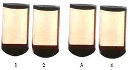
Figure 3: A visual picture condition of erythrocytes on 1st day.
Notes: 1 - control; 2 - after single processing by MCS-B; 3 - after double processing by MCS-B; 4 - after triple processing by MCS-B.
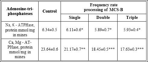
Table 3: Font results of research activity adenosinetriphosphatases before and after processing of erythrocytes by NPs of MCS-B (M±m; n=20).
Note: * - p>0.05; ** - p<0.01; *** - p<0.001
So, the dates of table 3 are demonstrating that single processing of blood by MCS-B reliably reduces (in comparison with the control) activity of Ca, Mg - АТPHase of erythrocytes - by 2.47±0.6 protein mmol/mg in mines (р<0.01), double - by 5.19±0.5 protein mmol/mg in mines (р<0.001), triple - by 6.01±0.5 protein mmol/mg in mines (р<0.001). On the contrary, the reliable changes activity of Na, K - АТPHase in any test tubes (in comparison with the control) were not detected (p>0.05).
It is known that if the enzyme level is reduced, this would lead to an increase in cytosolic calcium [19]. That is why cytosolic calcium was determined. Results of research the level of ion Ca2+ in erythrocytes before and after processing by NPs of MCS-B are presenting in table 4.
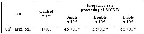
Table 4: Results research the level of ion Ca2+ in erythrocytes before and after processing by NPs of MCS-B (M±m; n=20).
Note: * - p<0.001 in comparative with control.
The dates of table 4 are demonstrating that level of cytosolic calcium with highly reliable (p<0.001) is increasing and depends from frequency rate processing by МСS-B.
It is known that increasing the concentration of cytosolic Ca2+ leads to a change in shape, decrease deformability, reduced life of erythrocytes and activation of eryptosis [20]. Also, increasing intracellular Ca2+ connected with increasing of erythrocytes aggregation and is the main reason of microcirculatory disorders [21]. If you follow the logic, in ours event the increase of cytosolic Ca2+ in erythrocytes must destroy them.
However, in this paper the results of studies reliably indicate the opposite. A visual condition of erythrocytes on stage II (21th day) is present on figure 4.
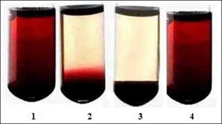
Figure 4: A visual picture condition of erythrocytes on 21th day.
Notes: 1 - control; 2 - after single processing by MCS-B; 3 - after double processing by MCS-B; 4 - after triple processing by MCS-B.
This picture is demonstrates that on stage II in tube of control the strongly pronounced sign of hemolysis was detected. Also, the strongly pronounced sign of hemolysis in tube of test after triple processing the blood by MCS-B was determined. In tube of test of blood that was single time processed by MCS-B the hemolysis was less pronounced. The sign of hemolysis in the tube of test after double processing the blood by MCS-B was not observed.
Thus, visual investigations have shown that maximum inhibition hemolysis of erythrocytes was determined after double processing by MCS-B. Opposite after triple processing of blood by MCS-B was not inhibited hemolysis of erythrocytes.
Represented the results of studies have shown that frequency rate processing of blood by MCS-B reliably influences on activity of hemolysis. However, appearance of hemolysis has not linear dependence on rise level of cytosolic calcium and frequency rate processing of blood by МСS-B. This does not contradict the mentioned before mechanisms, but only it proves multidirectional impact of nanoparticles MCS-B on the micro-cellular space, including the water sector, protein molecules and phospholipids. Maintaining membrane potential is necessary for normal functioning of ion channels that are very sensitive to any changes in them. Nanoparticles of MCS-B alter the bioelectric potential of erythrocyte membrane [22]. As a result, some ion channels open and ions including Ca2+ that is a regulator of many enzymes begin flow in cells according to concentration gradient. Therefore, increasing of intracellular concentration is a signal to start a series of processes such as synthesis of adenosinetriphosphate (universal cell “battery”) that is necessary for starting metabolic reaction [23]. The following sequence of events is: the action of a constant magnetic field of magnetite nanoparticles (300-400 kA/m) → membrane potential of cells changes → opening of ion channels, including calcium (Ca2+ begins to flow into the cell on the concentration gradient) → intracellular Ca2+ increases → activation of the Ca - depen- dent enzymes. Schematic illustration of the mechanism of the effect MCS-B on erythrocytes is present on figure 5.

Figure 5: Schematic illustration of MCS-B impacts on erythrocytes.
In result the energy appears that needs for further intracellular metabolic processes such as the activation of glycolysis in erythrocytes. As a result, 1.5 to 2 times oxygen capacity is increased, bioelectric charge membranes of erythrocytes are modulated catabolic processes in leukocytes are inhibited [23, 25] and eryptosis process is declining [19,23-25].
Thus, as a result of the research, the optimum frequency rate of extracorporeal processing of blood by NPs of MCS-B for maximum of slowing down of hemolysis was determined.
It was established, that extracorporally processing the blood by NPs of MCS-B reliably reduces activity of Ca, Mg - АТPHase of erythrocytes and increases level of cytosolic calcium.
This paper has shown that mechanism of inhibition hemolysis by NPs of MCS-B is not connects with increasing level of cytosolic Ca2+ and activity of adenosinetriphosphates (Ca, Mg - АТPHase).
Running a few steps forward next investigations have shown that activity of hemolysis depends on condition polarization of the water molecules micro-cellular space of erythrocytes. This is confirming Gilbert N. Ling’s theory about multi-layer organization polarization of water [26]. However, this scientific information will publish in next article.
Conclusion
- For inhibiting of hemolysis the optimum frequency rate (1-2 times) of processing the blood by NPs of MCS-B was determined
- It was established that extracorporally processing the blood by NPs of MCS-B reliably reduces activity of Ca, Mg - АТPHase of eryth- rocytes and increases level of cytosolic calcium
- After processing of blood by means NPs of MCS-B the activity of Na, K - АТPHase of erythrocytes does not change (p>0.05)
- Manifestation of hemolysis has not linear dependence on rise level of cytosolic calcium and frequency rate processing of blood by МСS-B
- Likely that the nanoparticles of MCS-B are changing the state polarization of water molecules of micro-cellular space of erythro- It is influences on activity of hemolysis, activity of АТPHases, opening of ion channels that in whole explains the decline of eryptosis mechanism.
Acknowledgment
The author is thankful to the department of biological of Kharkov National University and Kharkov Region Hospital.
References
- Lewinski N, Colvin V, Drezek, R (2008) Cytotoxicity of nanoparticles. Small 4: 26-49.
- Chen C, Li YF, Qu Y, Chai Z, Zhao Y (2013) Advanced nuclear analytical and related techniques for the growing challenges in nanotoxicology. Chem Soc Rev 42: 8266-8303.
- Pan Y, Du X, Zhao F, Xu B (2012) Magnetic nanoparticles for the manipulation of proteins and Chem Soc Rev 41: 2912-2942.
- Gao J, Gu H, Xu B (2009) Multifunctional magnetic nanoparticles: design, synthesis, and biomedical Acc Chem Re 42: 1097-1107.
- Reddy LH, Arias JL, Nicolas J, Couvreur P (2012) Magnetic nanoparticles: design and characterization, toxicity and biocompatibility, pharmaceutical and biomedical Chem Rev 112: 5818-5878.
- Mahmoudi M, Hofmann H, Rothen-Rutishauser B, Petri-Fink A (2012) Assessing the in vitro and in vivo toxicity of superparamagnetic iron oxide Chem Rev 112: 2323-2238.
- Wang Q, Shen M, Zhao T, Xu Y, Lin J (2015) Low toxicity and long circulation time of Polyampholyte-coated magnetic nanoparticles for blood pool contrast Sci Rep 5: 7774.
- Drašler B, Drobne D, Novak S, Valant J, Boljte S, et (2014) Effects of magnetic cobalt ferrite nanoparticles on biological and artificial lipid membranes. Int J Nanomedicine 9: 1559-1581.
- He Z, Liu J, Du L (2014) The unexpected effect of PEGylated gold nanoparticles on the primary function of Nanoscale 6: 9017-9024.
- Bankapur A, Barkur S, Chidangil S, Mathur D (2014) A Micro-Raman Study of Live, Single Red Blood Cells (RBCs) Treated with AgNO3 PLoS One 9: 103493.
- Kim MJ, Shin S (2014) Toxic effects of silver nanoparticles and nanowires on erythrocyte Food Chem Toxicol 67: 80-86.
- Watanabe M, Yoneda M, Morohashi A, Hori Y, Okamoto D, et al. (2013) Effects of Fe3O4 Magnetic Nanoparticles on A549 Cells. Int J Mol Sci 14: 15546-15560.
- Chen LQ, Fang L, Ling J, Ding CZ, Kang B, et al. (2015) Nanotoxicity of silver nanoparticles to red blood cells: size dependent adsorption, uptake, and hemolytic Chem Res Toxicol 28: 501-509.
- Belousov AN (1997) The method of producing magnetic liquid for transport and retention of medicines in the organism. The State patent №14817А UA A61N2/00/(Ukraine) Bull 2:3.
- Belousov AN (1998) The sorbent for extracorporeal detoxication of biological liquids: The State patent №24322А UA A61N2/00 / (Ukraine) Bull 7: 4.
- Belousov AN (2000) Micromage-B is product of treatment-and-prophylactic:The State patent №30538А UA A 23L 1/304/ (Ukraine) Bull 6: 3.
- Severina SE (1977) Transport Modern methods of exploration. Moscow State University Publishing house, Moscow, Russia.
- Belousov AN, Belousova E,Yu, Obolencev N (2005) Nanotechnology on way prolongation the vital process in organism. Pain, anesthetization and intensive - Kiev. - 2005. - №3. - 5-7.
- Belousov AN (2011) The Use of Magnetite Nanoparticles in Applied Medicine. Scientific.Net is a registered brand of Trans Tech Publications Inc 694: 205-208.
- Storozhok SA, Sannikov AG, Belkin AV (2009) Dependence of the stability of erythrocyte membrane deformability of intermolecular interactions of cytoskeletal Scientific Bulletin of the TGU - № 3. 3-10.
- Maymistova AA, Koshelev VB, Bulaeva SV (2010) Changing the aggregation and deformability of red blood cells in the activation of intracellular signaling Bulletin of the Yaroslavl Pedagogical № 3- 71-74.
- Zaguskni SL (2005) Intracellular mechanisms of laser MIS-RT.- № 36.
- Belousov AN (2004) Extracorporal hemocorrection using magnet-controlled sorbent in intensive therapy of intoxication syndromes in patients with hepatopancreatoduodenales diseases. Мanuscript. Dissertation for the Doctor of Medicine degree on speciality 14.01.30 - Anesthesiology and Intensive Dnepropetrovsk State Medical Academy.
- Belousov AN (2004) Influence of magnetite – preparation of nanotechnology on metabolism of cell. Bulletin of the problems of biology and medicine, Poltava,- №2. 34-37.
- http://www.techconnectworld.com/Biotech2012/sym/nano_medical_scienc- html
- Ling GN (2001) Life at the cell and below-cell level: the hidden history of a fundamental revolution in biology. Pacific Press, California, USA.
Citation: Belousov A, Belousova E (2017) Reducing of Erythrocytes Destruction By Means of Medicine Nanotechnology (Magnet-Controlled Sorbent Brand of MCS-B). J Cell Mol Biol 2: 005.
Copyright: © 2017 Belousov A. This is an open-access article distributed under the terms of the Creative Commons Attribution License, which permits unrestricted use, distribution, and reproduction in any medium, provided the original author and source are credited.