*Corresponding Author:
Andrey N Belousov,
Laboratory of Applied Nanotechnology of Belousov, Kharkov Medical Academy of Postgrad- uate Education , pr. Nauky, 31-v, fl 32, Kharkov, 61072, Ukraine
Tel: +380 509151889
E-mail: an.belousov2012@yandex.ua
Abstract
In an experiment it is shown on rats, that biocompatible standardized magnetite nanoparticle of ICNB can be effectively used at MRI. It is well proven that nanoparticles of ICNB for certain (р<0.001) strengthen a contrasting effect at MRI.
Methodology of safe intravenous application of ICNB is excluding the use of magnetite of nanoparticles in the variant of independent contrasting means at MRI. It is set that in 24 hours after intravenous injection of ICNB the magnetite of nanoparticles for certain (p<0.001) selectively accumulate in tissue of malignant tumour and rise brightness of image. In 96 hours after injection magnetite of nanoparticles the dynamics of reduction of brightness of image in tumour and muscles was establishment for certain (р<0.001). This fact is caused by process eliminating of nanoparticles from organism of rat. On the mechanism of action the nanoparticles of ICNB cause the convertible changes which is reason for the temporal increase of mobility of protons of hydrogen in near cell liquid. It inevitably modifies the metabolic process in malignant cells that in perspective has hope in elaborating new ways of the target therapy of malignant neoplasm.
Keywords
Contrast; ICNB; Magnetic; Malignant tumour; MRI; Nanoparticles; Selectively
Introduction
Idea of using magnetite of nanoparticles as contrasting means in MRI investigation is not new. Objectively it follows from physical properties of nanoparticles. Scientific literature abounds in information about the use of magnetite of nanoparticles as contrasting means [1-5]. Separate actuality application magnetite of nanoparticles has early MRI diagnostics and target therapy of malignant tumors. In spite of the fact that application of magnetite of nanoparticles looks simple, it is not necessary to forget about a high danger of the origins of complications as a result of their intravessel injection. At least it is necessary to take into account such indexes as a concentration, doze, rate of entered solution of nanoparticles, time of allocation nanoparticles in blood circulation after injection. The enumerated parameters for reliable have influence on haemorreology and state of microcirculation on the whole. The high local concentration of magnetite in vessels is caused by disturbances of blood circulation, microcirculation and hypoxia of tissues [6,7]. It is dangerous in main vital organs: brain, heart, lungs, liver and kidneys. Direct cross-correlation dependence between concentration of nanoparticles and level hypoxia is physiopathology obvious. Consequently, before injecting intravessel magnetite of nanoparticles, it is necessary to have standardized water solution magnetite of nanoparticles with early studied and well proven noninvasive physical and chemical properties.
Unfortunately, to date the advanced studies that would take into account it are absent. In the published advanced studies we met not a single reference to the use of the early studied standardized noninvasive forms magnetite of nanoparticles and methodologies of their application. Information about the mechanism of influence magnetite of nanoparticles on main biological systems of living organism including respiratory, cardiovascular, secretory, immune systems and cellular exchange is absent. Also we did not discover among the scientific publications of reliable dates about quantitative distribution magnetite of nanoparticles in organs and tissues after intravenous injection. Information about a mechanism of eliminate magnetite of nanoparticles from an organism is absent.
On the whole, aforesaid does not allow properly estimating the scientific and practicing significance early advanced studies which were published on theme to use magnetite of nanoparticles as contrasting means for MRI.
It was found in the choice of theme of the present investigation. The task was set in an experiment on animals to check possibility of the use of the before worked-out and studied methodology of intravenous injection of the standardized form water solution magnetite of nanoparticles (preparation of ICNB) [8-17] for contrasting of malignant tumour at MRI research.
The main purpose is to change the indexes of relaxation of T1 and T2 in area of malignant tumour during realization MRI by means of nanoparticles of ICNB.
Materials and Methods
Investigations were performed on the males of rats of vista line, by age of 26-27 months. Weight of rats was from 410 to 460 grams. Rats lived in individual cages with standard ration of vivarium with free access to water and food.
One rat was relatively healthy. Others are with the present adeno- carcinoma of mammary gland. During investigations of animals we observed the principles of humanity, which expounded in declaration of Helsinki.
For 5 minutes prior to research intramuscular the rats get seda- tion. Subsequently control of MRI was carried out.
After the performed control of MRI, singly in a tail vein of the rat, from a calculation 0.6-0.8 ml/100 mg, 0.0225% was injected ICNB. The repeated was performed of MRI studies. Conditionally all MRI studies were divided into 4 stages:
Stage I - is control (before intravenous injection of nanoparticles)
Stage II - in 5 minutes after injection magnetite of nanoparticles
Stage III - in 24 hours after injection magnetite of nanoparticles
Stage IV - in 96 hours after injection magnetite of nanoparticles (on 4th days)
Physical and chemical properties of ICNB (Figures 1&2; Tables 1&2)
- Osmolality theoretical of colloid solution is 500 mosm/l
- 0225% colloidal solution of magnetite nanoparticles
- Size of magnetite of nanoparticles is 6-12 nm
- Total area of surface magnetite of nanoparticles Ss = 800-1200 m2/g
- Magnetized of saturation is = 15 кА/m
- ζ - potential = - 19 mV

Figure 1: Study of magnetite nanoparticles (ICNB) with use microscope ion-elec- tronic raster-type Quanta 200 3 D.
The investigations were performed on the MR-tomagraph mag- neton concerto of siemens firm with power magnetic-field 0.2 T.

Figure 2: Study of magnetite nanoparticles (ICNB) with use microscope electronic translucent JEM-2100.
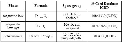
Table 1: X-ray analysis of ICNB in X-ray diffract meter Rigaku Ultima IV (CuKα, Kβ filter-Ni), one-coordinate Dtex semiconductor detector.
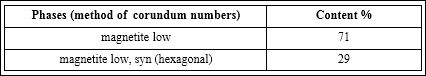
Table 2: The phases of ICNB (RIR-method; error 8±3%).
Got axial tomograms:
- T1-the self-weighted sequences of echo spin of TR 50 ms, TE 17 ms the field of review a 250 mm, the thickness of cut 2
- T2-the self-weighted sequences of echo gradient of TR 500 ms, TE 17 ms the field of review a 180 mm, the thickness of cut 4
The concentration of accumulation magnetite of nanoparticles was estimated by measuring brightness of image in a tumour and tis- sue of muscular of rats at MRI. The middle index brightness of image was accounted by measuring arbitrarily taken 8 points of minimum and maximal values of the investigated tissues. Got results were statis- tically processed by means of computer mathematical complex “stat- graf ”. The method of variation statistics comparison averages was used on the t-criterion of student.
Results and Discussion
Before the beginning implementation of MRI was performed re- search of ICNB for the purpose of visualizing his contrast effect. In parallel of solution 0.9% NaCl was studied for comparison. Results MRI of research ICNB and solution of 0.9% NaCl were presented in figure 3.
The figure 3 demonstrates the expressed contrasting effect of ICNB by comparing solution of 0.9% NaCl.

Figure 3: MRI research of ICNB and solution of 0.9% NaCl.
Initially the protons of atom of hydrogen in preparation of ICNB are in the maximally structured state and have low index of relaxation. Therefore, contrasting effect of ICNB at MRI is registered as darken- ing of image. This research confirmed possibility of the use of ICNB as contrasting means at MRI.
As a result of injection to the tail vein of rats preparation of ICNB at МРТ research an opposite effect is reliable. If figure 3 demonstrated the effect of darkening from ICNB at MRI, then after the intravenous injection of ICNB, opposite was exposed increase brightness of image in investigated tissues (Figure 4).
Author’s methodology the intravenous of injection allows to magnetite of nanoparticles of ICNB quickly dissolving in blood and in subsequent distributed in organs and tissues. Rapid dissolution of ICNB in blood prevents appearing rheological, microcirculation dis- orders and consequently the phenomena of hypoxia [6,7].
Distributed nanoparticles of ICNB in tissues against a back- ground of MR radiation strengthen influence of magnetic field on the protons of atom of hydrogen. The protons of atom of hydrogen alter the magnetic moment on opposite and then go back into initial position. As a result, energy increases in nucleus atoms of hydrogen, time of relaxation of the excited protons grows. It registers oneself the system of tomograph. The comparative image of contrasting effect be- fore and after injection of ICNB in rat with the adenocarcinoma of mammary gland is presented in figures 4 & 5.
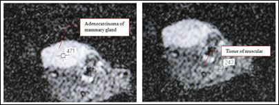
Figure 4: Initial MRI study the brightness of image in rat with the adenocarcinoma of mammary glandandtissue of muscular (471 conventional sign-tumour; 243 con- ventional sign-tissue of muscular).
Figures 4 & 5 in comparison show evidently that already on the first minutes after the intravenous injection nanoparticles of ICNB at MRI the parameters of relaxation T1 and T2 change and reliable (p<0.001) a contrasting effect increase in like brightness of image in the investigated tissues. So, after injection of ICNB in tumour of tissue the index of brightness of image increased on the average on 329±12 conventional sign and was 800±12 conventional sign (p<0.001), but in muscular on 457±12 conventional sign and was 700±12 conventional sign (p<0.001).

Figure 5: MRI study the brightness of image rat with the adenocarcinoma of mam- mary gland and tissue of muscular on the first minutes after intravenous injection of ICNB (800 conventional sign-tumour; 700 conventional sign-tissue of muscular).
It should be noted that registering the low parameters of re- laxation is possible only in case of high concentration magnetite of nanoparticles in blood and tissues. However, the high concentration of nanoparticles in blood stream is potentially dangerous for living organism, because it causes the origin of hypoxia in tissues. Especially this is very significant for organs: brain, heart, liver, lungs and kidneys.
Thus, taking into account foregoing, application magnetite of nanoparicles as contrasting means at MRI in the safe variant of meth- odology is practically not possible. This experiment showed that using nanoparticles of ICNB in certain methodology cannot be indepen- dent contrasting means at MRI. Worked out methodology of intra- venous injection nanoparticles of ICNB on a background of MR of radiation in it safe variant only for certain (p<0.001) strengthens the brightness of image of tissues. Dynamic change of indexes brightness image in tumour and muscular tissues in a sick rat after intravenous injection of ICNB is presented in figure 6. Dynamic change of indexes brightness image in muscular tissues in both rats groups after intrave- nous injection of ICNB is presented in figure 7.
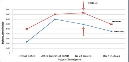
Figure 6: Dynamic change of indexes brightness image in tumour and muscular tissues in a sick rat after intravenous injection of ICNB on various stages at MRI investigation (n=8; p<0.001).
Opposite, in health rat on stage III investigation was reliable (p<0.001) revealed maximally increasing brightness of image in mus- cular tissue (700±19 conventional sing). Change of brightness image on stage III investigation in muscular tissue is caused by less accumu- lation in muscular tissue nanoparticles of ICNB in rat with tumour than in muscular tissue in health rat. This effect is explained as the following:
- The surface of malignant tumour cells as compared to healthy has higher negative charge, because nanoparticles of ICNB are selec- tively accumulated in malignant tumour tissue.
- Weak contact between cells of malignant tumour is reason for elevating accumulating nanoparticles of ICNB in intercellular space and rising of time of their elimination
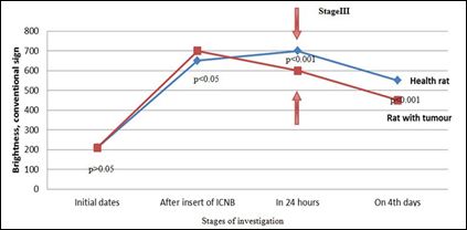
Figure 7: Dynamic change of indexes brightness image in muscular tissues in both rats groups after intravenous injection of ICNB on various stages at MRI investi- gation (n=8).
Weak contact between cells of malignant tumour is reason for elevating accumulating nanoparticles of ICNB in intercellular space and rising of time of their elimination
Conclusion
- In an experiment it is shown on rats that biocompatible standardized nanoparticles of ICNB can be effectively used at MRI. It is well proven that nanoparticles of ICNB for certain (р<0.001) strengthen a contrasting effect at MRI.
- Methodology of safe intravenous application of ICNB is excluding the use of magnetite of nanoparticles in the variant of independent contrasting means at MRI.
- It is set that in 24 hours after intravenous injection of ICNB the magnetite of nanoparticles for certain (p<0.001) selectively accumulate in tissue of malignant tumour and rise brightness of image.
- In 4th day investigation the dynamics of reduction of brightness of image in tumour and muscles was established for certain (р<0.001). This fact is caused by process eliminating of nanoparticles from organism of rat.
- On the mechanism of action the nanoparticles of ICNB cause the convertible changes which is reason for the temporal increase of mobility of protons of hydrogen in near cell liquid. It inevitably modifies the metabolic process in malignant cells that in perspective has hope in elaborating new ways of the target therapy of malignant neoplasm.
Acknowledgment
The author is thankful to the Department of Biological of Kharkov National University and department of MRI of Kharkov Region Hospital.
References
- Suzuki M, Honda H, Kobayashi T, Wakabayashi T, Yoshida J, et al. (1996) Development of a target-directed magnetic resonance contrast agent using monoclonal antibody-conjugated magnetic particles. Noshuyo Byori 13: 127- 132.
- Pankhurst QA, Connolly J, Jones SK, Dobson J (2003) Applications of mag- netic nanoparticles in biomedicine. J Phys D Appl Phys 36: 167-181.
- Salata OV (2004) Applications of nanoparticles in biology and Journal of Nanobiotechnology 2: 3.
- Bonnemain B (1998) Superparamagnetic agents in magnetic resonance imaging: physicochemical characteristics and clinical applications. A review. J Drug Target 6: 167-174.
- Weissleder R, Bogdanov A, Neuwelt EA, Papisov M (1995) Long-circulating iron oxides for MR imaging. Advanced Drug Delivery Reviews 16: 321-334.
- Patent of Ukraine №31309А UA A61N2/00 Method of treatment diseas- es which connected with blood circulation disturbance / A.N. Belousov № Decl. 04.08.98; Publ. 15.12.00.
- nanolab.com.ua
- Patent of Ukraine №14817А UA A61N2/00 Method of production of a mag- netic liquid for transport and retention of medicines in organism / A.N. Bel- ousov № 96062463. 21.06.96; Publ. 18.02.97.
- Patent of Ukraine №42123А UA A61N2/00 Method of treatment diseases which connected with endocrine disturbance / A.N. Belousov № 98084233. 04.08.98; Publ. 15.10.01.
- Patent of Ukraine 42132А UA A61N2/00 Method of treatment digestion system diseases / A.N. Belousov № 98084234. Decl. 04.08.98; Publ. 15.10.01.
- Belousov AN, Nevzorov VP (1998) Ultrastructure of Hepatic Cells in Rabbits after Injection of Magnetite Nanoparticles. Eighth International Conference on magnetic fluids 482-483.
- Belousov AN, Rykov VG (1998) Revealing of mechanisms of the influence exerted by magnetic fluid preparations on biological systems and the whole living organism.
- Belousov AN (2000) Experimental study of effects of Belousov’s mag- net-controlled sorbent on parameters of acid-base equilibrium in blood and processes of glycolysis in erythrocytes. Adsorption technologies and blood purification procedures, Germany.
- Belousov AN (2008) Opening the mechanisms of cell regulation by nanotechnology preparations. /7th International Conference on the Scientific and Clinical Applications of Magnetic Carriers - Vancouver, Canada.
- Belousov AN (2012) Effect magnetite nanoparticles MCS-B on functional activity of erythrocytes / ICEEP 2012: Electrical Power & Energy Systems. Hohhot, China.
- Belousov AN (2011) The influence of magnetite nanoparticles (MCS-B) on the hemolysis of erythrocytes / World Conference and Expo 2011. Tech- Connect World is the host of the Nanotech, BioNanotech, Microtech, Clean Technology and TechConnect conferences. June 13-16, 2011, in Boston, Massachusetts, USA.
- Belousov AN (2000) Application of products nanotechnology at creation of devices “An Artificial Liver”. / 12-th World Congress of Anesthesiologists, Monreal, Canada.
Citation: Belousov AN (2017) New Prospects Application of Magnetite Nanoparticles for the Diagnosis of Malignant Tumors in MRI Investigation. J Cell Mol Biol 2: 003.
Copyright: © 2017 Belousov AN, et al. This is an open-access article distributed under the terms of the Creative Commons Attribution License, which permits unrestricted use, distribution, and reproduction in any medium, provided the original author and source are credited.