*Corresponding Author:
Sana Mallouk,
Department of Otorhinolaryngology, Head and Neck Surgery, King Université Hassan II, Casablanca, Morocco
Tel: +212 660729971
Email: sanaa.mallouk@gmail.com
Abstract
Tuberculosis is one of the world’s leading causes of death. Laryngeal tuberculosis is one of the extrapulmonary tuberculosis affecting different parts of the larynx with or without pulmonary involvement. Laryngeal carcinoma, which shares almost the same symptoms and signs, should be ruled out immediately. We report a case of laryngeal tuberculosis, initially misdiagnosed as laryngeal carcinoma; A 57 years old man, with history of smoking. Suspended laryngoscopy examination revealed the presence of a budding mass at the epiglottis, so the first diagnostic was laryngeal malignancy. Histopathological examination of the laryngeal lesions revealed chronic granulomatous inflammation with caseous necrosis without any signs of malignancies.
Tuberculosis and carcinoma of the larynx can have many clinical similarity, moreover it is important to distinguish between them, as a delay in diagnosis or improper diagnosis can significantly alter the management plan.
Keywords
Laryngeal malignancies; Laryngeal tuberculosis; Tuberculosis
Introduction
Tuberculosis is one of the world’s leading causes of death due to an infectious agent, and remains a major public health burden in many developing countries, 1 with 9.2 million new cases and 1.7 million deaths expected every year [1]. TB affects people of both sexes in all age groups but the highest burden is in adult men, who accounted for 57% of all TB cases in 2018. By comparison, adult women accounted for 32% and children for 11%. Among all TB cases, 8.6% were people living with HIV [2].
Laryngeal tuberculosis is one of the extrapulmonary tuberculosis affecting different parts of the larynx with or without pulmonary involvement [3]. Awareness of the disease is important, as it is frequently misdiagnosed as a malignant laryngeal tumor or granulomatous disease [4]. We report a case of laryngeal tuberculosis, initially misdiagnosed as laryngeal carcinoma.
Case Report
A 57 years old man, with history of smoking, was admited to our departement for permanent chronic dysphonia evolving for 6 months without dyspnea or dysphagia, the patient report also an 8 KG weight loss during the past 10 month. There was no history of any medication use or allergies.
The CT scan of the neck showed a regular thickening of the epiglottis (Figure 1). On the other hand, the glotto-susglottic region is without abnormalities. Suspended laryngoscopy revealed the presence of a budding mass at the lingual face of theepiglottis, associated with multiple leukoplakic lesions in the glotto-susglotticregion, especially in the two vocal cords, the two ventricular bands and the two arrhythenoids. Multiple biobsies have been realized. Histopathological examination of the laryngeal lesions revealed caseo-follicular tuberculosis laryngitis.
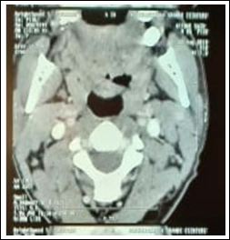
Figure 1: Axial contrast-enhanced CT of the larynx shows regular thickening of the epiglottis
In the search for primary tuberculosis, a chest X-ray shows peripheral opacity upper left lobe (Figure 2). The bacteriological examination found resistant acid-alcohol bacilli.
On re-questioning the patient at this stage, he admitted to a mild nocturnal cough, and some night sweats. The patient have received antibacillary treatment based on 6-month regimen of Isoniazid (INH), Rifampin (RIF), Pyrazinamide (PZA) and Ethambutol (EMB) for an intensive phase of 2 months, followed by a continuation phase with 4 months treatment of INH and RIF.
During the follow-up, 4 months after starting treatment, the patient reported an important clinical improvement, and endoscopic control showed a clear regression of the laryngeal lesions.
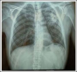
Figure 2: Initial antero-posterior chest X-ray shows peripheral opacity upper left lobe.
Discussion
Tuberculosis (TB) remains a major cause of ill health and is one of the top 10 causes of death worldwide. An estimated 10.0 million (range, 9.0-11.1 million) people fell ill with TB in 2018, a number that has been relatively stable in recent years [2]. Morocco has had a national tuberculosis control programme for more than 40 years, and was one of the first countries to adopt the DOTS (directly observed treatment, short-course) strategy in the early 1990s. For the past 10 years, the Moroccan National Tuberculosis Programme (NTP) is estimated to have met and exceeded international targets for estimated case detection and treatment success rates [5].
The clinical pattern of presentation of laryngeal tuberculosis has changed over the years. Formerly, laryngeal involvement was always associated with advanced pulmonary infection with constitutional symptoms of productive cough, fever, weight loss, hemoptysis and change in voice [3].
A metanalyse made by Xu Qian and coleagues, have found that, larynx is the second most common site of manifestations for head and neck tuberculosis, and Laryngeal lesions account for approximately 1% of TB cases found in the head and neck area [6].
Laryngeal tuberculosis was almost always associated with advanced pulmonary infections. However, recent cases reports have described a laryngeal tuberculosis with negative findings on radiological examinations of the chest and negative sputum cultures [7-9]. In our case we think that the laryngeal tuberculosis is secondary to the pulmonary infection.
JEANA L and al. [10] have undertaken a systematic review of published cases in the USA. They found that laryngeal tuberculosis was commonly localized at vocal cords; the true vocal cords were involved in 25 cases, false vocal cords in 15, the remaining cases involved the epiglottis in 13, larynx in 5, arytenoids in 5, interarytenoid in 4, aryepiglottic fold in 2, and subglottic/ cricoarytenoid in 2. The type of involvement varied, with 26 cases described as exophytic or mass lesion, 16 edema or erythema, 10 ulcerative, 6 polypoid, 5 nodular, 3 granular, and 5 other or non-specified. Tuberculosis and carcinoma of the larynx can have much clinical similarity, such as, dysphonia, dysphagia, wight loss and history of smoking, moreover it’s too difficult to differentiate laryngeal lesions on endoscopy.
Laryngeal TB manifested as a diffuse bilateral lesion with or without a focal mass and was always associated with active pulmonary TB. Although the CT appearances may not be specific, the possibility of TB should be raised when a bilateral and diffuse laryngeal lesion is seen without destruction of the laryngeal architecture in patients with pulmonamy TB [10,11]. It is important to distinguish between LB and malignancies, Tuberculosis is amenable to medical management, whereas, malignancy warrants surgery to the extent of total laryngectomy. Our case is a typical example, because of the history of heavy smoking and presence of budding mass of epiglottis, we had not considered TB until the histopathological result who showed caseating necrosis, which is consistent with tuberculosis infection.
Laryngeal TB is a rare differential diagnosis to be considered in cases presenting with clinical and laryngoscopic findings of malignancy, but should be suspected as a delay in diagnosis or improper
References
- Tachfouti N, Nejjari C, Benjelloun MC, Berraho M, Elfakir S, et al. (2011) Association between smoking status, other factors and tu- berculosis treatment failure in Morocco. Int J Tuberc Lung Dis 15: 838-843.
- World Health Organization (2019) Global tuberculosis report 2019. World Health Organisation, Geneva , Switzerland.
- Lynrah KG, Tiewsoh I, Marbaniang E, Barman B, Synrem E, et al. (2018) Laryngeal tuberculosis not uncommon in the present era. J Tuberc Ther 3: 118.
- Richter B, Fradis M, Köhler G, Ridder GJ (2001) Epiglottic tubercu- losis: Differential diagnosis and treatment. Case report and review of the Ann Otol Rhinol Laryngol 110: 197-201.
- Ottmani S, Obermeyer Z, Bencheikh N, Mahjour J (2008) Knowledge, attitudes and beliefs about tuberculosis in urban Mo- East Mediterr Health J 14: 298-304.
- Qian X, Albers AE, Nguyen DTM, Dong Y, Zhang Y, et al. (2019) Head and neck tuberculosis: Literature review and meta-analysis. Tuberculosis (Edinb) 116: 78-88.
- Huon LK, Fang TY (2011) Primary laryngeal J Formos Med Assoc 110: 792-793.
- Edizer DT, Karaman E, Mercan H, Alimoglu Y, Esen T, et (2010) Primary tuberculosis involving epiglottis: A rare case report. Dys- phagia 25: 258-260.
- Agarwal R, Gupta L, Singh M, Yashaswini N, Saxena A, et (2019) Primary laryngeal tuberculosis: A series of 15 cases. Head Neck Pathol 13: 339-343.
- Benwill JL, Sarria JC (2014) Laryngeal tuberculosis in the United States of America: A forgotten disease. Scand J Infect Dis 46: 241-249.
- Moon WK, Han MH, Chang KH, Kim HJ, Im JG, et al. (1996) La- ryngeal tuberculosis: CT findings. Am J Roentgenol 166: 445-449.
Citation:Mallouk S, Radi S, Baghdad M, Oukessou Y, Rouadi S, et al. (2020) Laryngeal Tuberculosis Mimicking Supraglottic Carcinoma: A Case Report and Review of Litera- ture. J Case Repo Imag 4: 016.
Copyright: © 2020 Mallouk S, et al. This is an open-access article distributed under the terms of the Creative Commons Attribution License, which permits unrestricted use, distribution, and reproduction in any medium, provided the original author and source are credited.
