*Corresponding Author:
Gunes Orman,
Department of Otorhinolaryngology, Head and Neck Surgery, King Université Hassan II, Casablanca, Morocco
Tel: +212 664689902
Email: mohamedbeghdad13@gmail.com
Abstract
Epidermal cysts are common skin lesions. There are only few case reports of epidermal cyst in submandibular gland that have been reported in the literature. Histopathological examination is required for confirmation of diagnosis. We report here a rare case of an epidermal cyst in the right submandibular gland in a 21 years old man patient.
Keywords
Epidermal cyst; Submandibular gland; Surgery
Introduction
Epidermal cysts are a benign lesion usually found in the skin and that are derived from abnormally situated ectodermal tissue. Salivary gland location is a rare entity. They usually become apparent in patients between 15 to 35 years. The diagnosis of an epidermal cyst in the submandibular gland becomes very essential and it could be mistaken for a salivary gland abscess, neoplasm, tuberculosis lymphadenitis, metastatic node or mucocele. They can cause symptoms of dysphagia and dyspnea. Diagnosis is difficult using imaging and clinical findings alone. Histopathological examination of the cyst is required for confirmation of diagnosis.
Case Report
A 21 years old male patient without any particular pathological history of trauma or previous facial surgery was admitted to our ENT department with a six years history of a progressive swelling on the right submandibular region, with difficulty in swallowing for 6months. There was no history of pain, fever, or any discharge from the lesion.
Initial examination showed a large mass measuring 5×3 cm in dimension in the right submandibular region. On palpation, the swelling was painless, mobile, and firm. There was no enlargement of the floor of the oral cavity and the duct of the right submandibular gland was normal. There was no palpable cervical lymphadenopathy.
A cervical Computed Tomography (CT) scan showed a weld limited oval hypodense lesion of the right submandibular region causing a digestive repression and measuring approximatively 56×31×28 mm (Figure 1).
We performed a total excision of the cyst under general anesthesia. A 4 cm line extending anteriorly from the anterior border of the sternocleidomastoid muscle and parallel to the mandible was marked at 2 fingers below the angle of mandible. Mandibular nerve was preserved. A cystic lesion measuring 6×3 cm was totally removed. A portion of submandibular salivary gland was also removed, attached posteriorly to the cystic swelling (Figure 2).
Histopathological examinationrevealedstratifiedsquamous epithelium with an intraluminal laminated keratinized material confirming the diagnosis of epidermal cyst in the right submandibular gland.
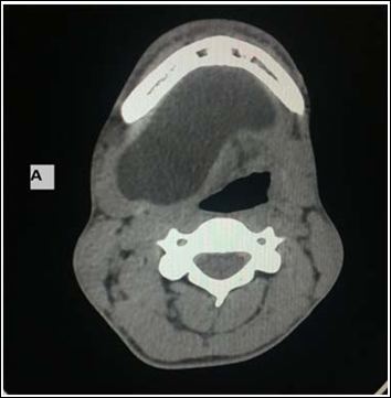
Figure 1: Cervical CT scan: Well limited cyst lesion of the right submandibular region causing a digestive repression.
A/ axial view. B/ coronal view
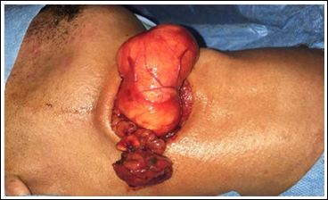
Figure 2: Excision of the total epidermal cyst attached to the right submandibular gland.
Post operatively, we did not notice any abnormalities, and after 2 days, the drain was removed then the patient was discharged. In¬ 6 months follow up, there were no signs of recurrence.
Discussion
Epidermal cysts are very common skin lesions also known as epithelial or infundibular cysts, and it is a distinct pathological entity from the epidermoid cyst [1].
They appear as intradermal or subcutaneous tumors with a progressive development on the face, neck, back and scrotum. These lesions usually occur secondary to obstruction, lined by stratified squamous epithelium and filled with laminated keratin, cholesterol crystals, and debris [2].
Epidermal cyst of the submandibular gland are relatively uncommon and is likely to be mistaken for a salivary gland abscess, neoplasm, TB lymphadenitis, metastatic lymph node or any other cysts [3].
The clinical and radiological differential diagnosis of cystic lesions of the submandibular region can be difficult. Patients usually have a swelling of the submandibular region that gradually increases in size with no inflammatory signs. If the cyst stays for a longer period of time, it might get infected forming fistulas. It might also give compression symptoms like dysphagia, dyspnea or dysphonia [4].
Histologically epidermal cyst has stratified squamous epithelial lining and is usually filled with keratinized material [5].
Complete local excision of the cyst without any rupture is considered to be the best treatment .The recurrence rate of an epidermal cyst is low [6]. In our case, there has been no recurrence in 6 months follow up.
Conclusion
Epidermal cysts of the submandibular gland are rare. Best treatment option for such cases is local complete excision of cyst to confirm diagnosis and to prevent complications due to cyst growth.
References
- Ganesan A, Nandakumar GK (2015) Epidermal cyst of parotid gland: A rarity and a diagnostic Case Rep Dent 2015.
- Baschinsky D, Hameed A, Keyhani-Rofagha S (1999) Fine-needle aspiration cytological features of dermoid cyst of the parotid gland: A report of two cases. Diagn Cytopathol 20: 387-388.
- Dutt SN, Hock YL, Saleem Y, Bhat N, East DM (2000) Epidermoid Cyst of the Submandibular Gland. Indian Journal of Otolaryngolo- gy& Head &Neck Surgery 52: 378-379.
- Kar R, Thorawade V, Jagade M, Attakkil A, Kedar S, et (2014) An Unusal case of epidermal cyst in submandibular space. Internation- al Journal of Otolaryngology and Head & Neck Surgery 3: 213-215.
- Janarthanam J, Mahadevan S (2012) Epidermoid cyst of subman- dibular J Oral Maxillofac Pathol 16: 435-437.
- Patil P, Shukla D, Sonawane R (2016) A rare case of epidermal cyst of the submandibular gland. International Journal of Health Scienc- es & Research 6.
Citation:Beghdad M, Laachoubi M, Choukry K, Mkhatri A, Oukessou Y, et al. (2020) Epidermal Cyst of the Submandibular Gland: A Case Report. J Case Repo Imag 4: 017.
Copyright: © 2020 Beghdad M, et al. This is an open-access article distributed under the terms of the Creative Commons Attribution License, which permits unrestricted use, distribution, and reproduction in any medium, provided the original author and source are credited.
