*Corresponding Author:
Andrea Pirri,
Emergency Department, ARNAS G. Brotzu, Cagliari Italy
E-mail: andreapirri@tiscali.it; andreapirri@aob.it
Abstract
A 45-year-old man trasported for syncope and persistent altered state of consciouness to the Emergency Department (E.D) of A.R.N.A.S G. Brotzu in Cagliari (Italy). The patient presented a state of agitation, bilateral pupillary myosis and hyposthenia of the right part of the body, we performed a head CT scan that showed “minimal signs of subarachnoid hemorrhage in the left fronto-parietal. Subsequently pallor of the feet with absence of palpation of the pedid pulses appeared for this reason we performed a ecocardiogram that showed a large oblong mobile mass that occupied half of the atrium, a chest and abdomen CT angiography showed an image of “minus” in the left atrium , subsequently the patient underwent an operation to unblock the aorto-bisiliac axis with a Fogarty catheter and cardiac surgery to excise the atrial myxoma, despite the early two interventions the patient had a poor prognosis and remained in a post anoxic state of coma.
Keywords
Atrial myxoma; Clinical presentation; Outcome
Introduction
A 45-year-old man is rescued in February 2019 by a medical crew of Territorial Emergency System in Cagliari (Sardinian island, Italy) for a syncopal episode with head injury and altered state of consciousness.
Upon arrival of medical crew, the patient was confused, presented with bilateral pupillary myosis and right brachio-crural weakness [1]. After direct assistance to the patient, the medical crew consulted the patient’s wife who told them he had any illness and not used illicit substances , but two days ago the husband had complained for a transient chest disconfort. The patient’s haemodynamic parameters were within normal limits, because the physician suspected a possibile opiate overdose he adiministered to the patient 3 vials of naloxone with transient resolution of myosis and transported him to the Emergency Department (E.D) of A.R.N.A.S G. Brotzu in Cagliari (High Specialty Hospital) .
In E.D the neurological state of man was substantially unchanged (GSC 8 : E2V2M6) the patient presented a state of agitation, bilateral pupillary myosis and hyposthenia of the right of the body , the vital signs were unchanged except a minor desaturation (Spo2 91% in ambient air ), we performed various tests including: electrocardiogram, blood exames , ABG (PH 7.4, PCO2 32 mmHg, PO2 140 mmHg, glycemia 128 mg /dl, LAT 2.1), a consultation of a resuscitator physician, a Point-of-Care ultrasonography which was difficult to perform due to the patient’s agitation, did not show pleural, pericardial and free abdominal effusion, no dilatation or dissection images of the first tract of the thoracic ascending aorta and abdominal aorta. Subsequently we performed a head CT scan (Figure 1) which showed “minimal signs of subarachnoid hemorrhage in the left fronto-parietal without evidence of abnormal vascular structures” and after neurosurgical evaluation the patient was admitted to the Stroke Unit and because of the signs of cerebral haemorrhage he was not subjected to intravenous thrombolytic therapy.

Figure 1: Head CT scan (“minimal signs of ESA in the left fronto-parietal”).
Upon entering the Stroke Unit, the neurological state was further compromised (GCS 7 E1V1M5: motor response 5 for the left upper limb, no motor response to the pressure stimulus in the right upper limb and lower limbs), pallor of the feet with absence of palpation of the pedid pulses, for this reason a vascular surgical consultation with echo-color doppler of the arterial circulation of the lower limbs was requested which highlighted an “acute bilateral ischemia of the lower limbs with absence of femoral pulses” for this reason chest and abdomen CT angiography was required. While waiting to perform the aforementioned examination, an echocardiogram (Figure 2) was requested which showed a large oblong mobile mass that occupied half of the atrium with dimensions of 80x30 mm with a pedunculated appearance with a narrow implant base on the lateral wall and with fringed and irregular surface, very mobile, which during diastole engaged through the mitral valve even if apparently only partially oc- cluding its opening, no evidence of contact points nor adherence with the mitral flaps, left atrium of normal size, no evidence of pericardial effusion, apparently tricuspid aorta, aortic bulb of normal size.

Figure 2: Sub xyphoid echocardiography scan.
The chest and abdomen CT angiography (Figure 3) performed immediately afterwards showed a coarse image of “minus” in the left atrium (max diam. 8 cm) in probable relationship with the mitral valve, thrombotic occlusion of the abdominal aorta at the level of the carrefour extended to both iliac axes up to at the bifurcation, multiple bilateral splenic and renal infarcts [1]. During the night of hospitalization the patient underwent an operation to unblock the aorto-bisiliac axis with a Fogarty catheter by the Vascular Surgery team and the next day he underwent a cardiac surgery to excise the atrial myxoma and subsequently transferred from the Cardioanesthesia Division to the Intensive Care Unit (I.C.U) of our Hospital. The hospitalization in (I.C.U) was complicated by the formation of a pericardial effusion subsequently drained by the cardiac surgeon, the clinical conditions and neurological state of post-ischemic coma required the placement of a tracheostomy and a Percutaneous Endoscopic Gastrostomy (PEG); on 2 April 2019 he was transferred to a Teritorial Unit for the rehabilitation treatment of the case.
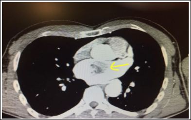
Figure 3: Chest CT angiography (cardiac minus image).
Discussion
Atrial myxomas [1] are the most frequent primary cardiac tumors, due to non-specific symptoms they can have a late diagnosis, they represent 40-50% of primary heart tumors, 90% are solitary and pedunculated and in 75-85% of cases they are located in the left atrium; in 10% of cases they occur in a familial form and are transmitted as an autosomal dominant trait. From a macroscopic point of view they have a gelatinous consistency and a yellow / brown color with a mammellon surface while from a histological point of view they are of a stromal and vascular nature and can be fed by a coronary branch, in the benign forms they can recur if the excision is incomplete and rarely have a malignant character and embolize (metastatic emboli) even at a distance. The dimensions can vary from 1 to 15 cm in diameter.
The onset symptoms are related to the haemodynamic consequences on cardiac function and on the mitral and tricuspid valve, for the possible prolapse in the ventricle in polypoid forms and for the thrombo-embolic phenomena that are generated at a distance. The sites of embolism depend on the site of the tumor (right or left atrium), and are sometimes associated with aneurysms in the lung. The main complications are: heart failure, cardiac arrhythmias, myocardial infarction, sudden death.
In 20% myxomas of cases may not give to be relevant symptoms and be diagnosed accidentally during the execution of an echocardiogram, in other cases there are signs and symptoms of stenosis and regurgitation of the mitral valve, signs of left heart failure (exertional dyspnea, orthopnea, pulmonary edema), signs of right heart failure (asthenia, peripheral edema, rarely ascites), dizziness, syncope (20% of cases). endocarditis or signs and symptoms of a collagen disease.
Myxoma-related embolism can produce transient ischemic attacks, strokes or seizures in the central nervous system, there may be signs and symptoms related to pulmonary or systemic embolism depending on the location of the tumor in the right or left atrium or depending on the presence of shunts associated cardiac disorders . In a review of 113 cases of patients with atrial myxoma and onset neurological symptoms [1], 83% of patients presented with ischemic stroke in various locations (in 43%) and 20% with epileptic seizures. systemic embolism in any artery (coronary, aorta, renal, visceral or peripheral) can cause heart attacks or signs of ischemia in the corresponding organs and sites [2]. In the case of right atrial myxoma microembolisms can cause pulmonary hypertension and chronic pulmonary heart, general symptoms may be fever, weight loss, arthralgia. Infrequent, a atrial myxoma the latter can be linked to coronary embolism, while endocarditis is a consequence of the formation of vegetations on the surface of the tumor. On physical examination, jugular turgor may be found, a third S1 tone due to delayed mitral valve closure due to tumor prolapse in the valvular plane, a diastolic murmur may be heard due to the impact of the tumor against the endocardial wall, or due to obstruction of the mitral or tricuspid valve plane. In Camey’s syndrome, myxoma is associated with multiple brain aneurysms.
The exames of choice in the diagnosis are the Trans-thoracic (TTE) and Trans-esophageal (TEE) echocardiogram, the TTE allows to evaluate the size, location, number of myxomas, the presence of peduncle and its mobility and hemodynamic consequences with respect to the valve planes. TEE has better specificity and 100% sensitivity than TTE.
The CHEST X-ray: the chest X-ray can only show an alteration of the cardiac profile. The CT scan of chest not only allows a study of coronary arteries and cardiac structures but allows a differential diagnosis with respect to intracardiac thrombi that have a narrower implant base and compared to other cardiac pathologies. The magnetic resonance (MRI) can better characterize site, size, shape, implant base and post-surgical correlation.
It’s important make differential diagnosis towards intracardiac thrombosis and metastatic (secondary) cardiac tumors, mitral insufficiency / stenosis, tricuspid insufficiency / stenosis, idiopathic pulmonary hypertension. The cardiac catheterization can be useful in the preoperative phase to evaluate tumor neovascularization and the concomitant existence of atherothrombotic coronary disease in patients over 40 years of age. The treatment of a atrial myxoma is the its excision and the post-operative mortality was found in 2.2% of cases.
References
- https://www.uptodate.com/contents/cardiac-tumors
- Ali MU, Finkel J (2018) Images in Clinical Medicine Atrial Myxoma. N Engl J Medm 379: e26.
Citation: Pirri A, Telmon MC, Polo F (2021) Dramatic Clinical Presentation of Atrial Myxoma. J Emerg Med Trauma Surg Care 3: 012.
Copyright: © 2021 Pirri A, et al. This is an open-access article dis- tributed under the terms of the Creative Commons Attribution License, which permits unrestricted use, distribution, and re- production in any medium, provided the original author and source are credited.