*Corresponding Author:
Omar Iziki,
Department of Otorhinolaryngology, Head and Neck Surgery, King Université Hassan II, Casablanca, Morocco
Tel: +212 0613953181
Email: Omar.iziki@gmail.com
Abstract
The concha bullosa is the most common anatomic variation of the middle turbinate of the paranasal sinuses. As a result, the middle meatus get narrowed by the pneumatization of a turbinate, blocking the sinus draining, and its mucosal secretion leads to a mucocele that transforms to a pyocele after the liquid infection. We report a rare case of an adolescent with a pyocele of concha bullosa revealed by an intranasal mass.
Introduction
The concha bullosa is a common anatomic variant, represented by the pneumatization of a turbinate, usually the middle turbinate. Frequently asymptomatic, the obstruction of its ostium may lead to a mucocele that evolves to apyocele after infection of retained secretion. It’s a rare condition that appears as nasal mass, possibly mistaken for a neoplasm. The diagnosis is considerably helped by the radiological signs, while the confirmation is based on the surgical exploration.
Case Report
We report the case of an 18 years old girl, with no medical history, who suffered from a left permanent nasal obstruction for a year. The clinical examination found a large nasal mass with no other rhinological sign.
The facial CT scan showed a voluminous and heterogeneous polypoid leftnasal process with calcifications, measuring approximately 58mm x 25.4mm, expanded on 36mm, arriving posteriorly to the ipsilateral choana, invading the right ethmoidal labyrinth with partial bone erosion, till the nasal septum which is repressed with partial lysis of its bony portion; laterally, the process attains the medial wall of the right maxillary sinus, causing sinus retention (Figure 1).
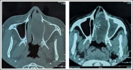
Figure 1: (A) CXR showed elevated left hemidiaphragm with bowel loop suspecting diaphragmatic hernia. (B) CT axial image confirming part of bowel herniated into the left thoracic cavity.
The treatment was based on a total surgical removal under general anesthesia. During the surgery, was discovered a voluminous mass, with a regular aspect, on the left middle turbinate, going continuously with its mucosa. The structure was opened exposing a cavity into the middle turbinate with pus flowing. The instrumental excision of the mass was performed with a middle meatotomy, a total ethmoidectomy and a right sphenoidotomy, in order to permit the sinus draining (Figure 2)
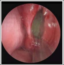
Figure 2: 3D endoscopic image showing the concha bullosa opened exposing a cavity into the middle turbinate with pus flowing.
Discussion
The concha bullosa is the most frequent accessory sinus pneumatisation, estimated at more of 30%, possibly concerning all the turbinates, mostly the middle one in 14 to 15% of the population. In this case, the pneumatization results from an extension of the anterior (55%) or posterior (45%) ethmoidal cells [1,2]. As a result, the middle meatus get narrowed blocking the sinus draining, and its mucosal secretion leads to a mucocele that transforms to a pyocele after the liquid infection.
Until today, little is known about the pathogenesis of middle turbinate concha bullosa mucocele. It is believed that they form due to chronic blockage of the concha bullosa ostium, which prevents appropriate airflow between the pneumatized cavity and its originating compartment. Thus, it could explain why the epithelial contour remains intact in mucoceles of sinonasal compartments. This is in contrast to the conventional mucocele definition that refers to a trauma-induced, fluid-filled cavity that lacks an epithelial surrounding. Previous studies indicate either a mechanical (history of trauma, surgery, nasal polyposis, or benign tumors) or an inflammatory etiology (infection, allergy, cystic fibrosis), whereas others did not support the obstructive causes of mucoceles. Otherwise, it can be mistaken for neoplasms [2]. In our case, no specific medical history was found, an absence of nasal trauma or rhinosinusitis.
The anterior rhinoscopy is an essential method of nasal cavity examination, even if it is not appropriate for evaluating a concha bullosa or a mucopyocele, it helps to appreciate the size and degree of nasal airway obstruction [3]. The clinical findings may vary. The most common symptoms are the nasal mass or middle turbinate hypertrophy, like in our case, with or without septal deviation. If the mucosal lining of pneumatized middle turbinate becomes inflamed, symptoms such as nasal obstruction, post-nasal discharge, snoring, headache, and fever occur [4]. The gradual mass effect and the fact that a mucocele in a concha bullosa has more surrounding space to expand can explain the lack of prevalent signs even with a large size mass and then, the possible diagnosis delay [2]. However, a direct extension into the orbit may occur, associated with ocular functional loss, because the orbital contents are separated from the ethmoidal labyrinth only by the thin lamina papyracea. In some cases, the ocular motion and vision remains normal despite of the presence of orbital extension for years [5].
The facial CT scan is the ideal imaging technique, offering a perfect visualization of the concha bullosa and a clear detection of its mucocele. But, it can’t differentiate between a mucocele and a pyocele. They both appear as a soft-tissue mass with a bony contour, usually causing deviation or compression of surrounding structures. The bony rim is considered the main finding on CT that enables identification of the mucocele into the middle turbinate; if absent, the diagnosis may be confused with a tumour, then an incisional biopsy can be done [2,3]. The absence or inconsistent presence of bony shell could be explained by an underlying mechanism of bone remodelling [2]. Also, a mucopyocele can be suggested in the presence of peripheral enhancement of a mucocele, as shown in our case [3].
On another hand, the MRI is useful to detect the extranasal extension, evaluate the vascularity of the mass and delineate secretions. Thereby, it can differenciate between a concha bullosa mucopyocele and a tumour which enhaces with gadolinium [2]. The MRI wasn’t performed in our patient, because of its cost that may lead to a therapeutical delay.
Concerning the microbiology results of culture of the retention liquid, S. aureus is the most common organism seen in the literature, with no reports of Corynebacterium species, E. coli, or H. influenza, and only one described case of fungi [2]. Unfortunately, no analysis was done on the pus in our case.
The endoscopic nasal surgery is the favoured treatment, based on a transverse excision, a crushing, or a lateral or medial marsupialization. In our case, after the opening of the mass confirming its nature, the marsupialisation was performed followed by an enlargement of the sinuses open which is considered providing the highest rate of success for these surgeries. In addition, no change in surgical procedure is necessary for patients with orbital involvement because decompression and resolution of orbital symptoms occur almost immediately after surgery [2].
The histological examination of the mass usually reveals an active chronic inflammation of a respiratory mucosa. The inflammation can be secondary to a foreign body [4].
Conclusion
To conclude, the concha bullosa is the most frequent accessory sinus pneumatisation, that can, in rare instances, develop a mucocele or, if infected, a pyocele. The major symptom is a nasal obstruction with a mass visualized on the anterior rhinoscopy. Thus, although a rare entity, it is important to consider the concha bullosa mycocele or pyocele in the differential diagnosis of nasal masses. The CT scan permits a precise analysis of the mucocele, even without detecting the difference with a pyocele. If symptomatic, an endoscopic surgery is performed consisting on the excision or marsupialization of the mass with widening the blocked sinuses draining sites.
References
- Marsot-Dupuchet K, Genty E (2003) Les variantes anatomiques des sinus de la face. Journal de radiologie 84: 357-367.
- Khalife S, Marchica C, Zawawi F, Daniel SJ, Manoukian JJ, et al. (2016) Concha bullosa mucocele: A case series and review of the Allergy Rhinol (Providence) 7: 233-243.
- Al-Sebeih KH, Bu-Abbas MH (2014) Concha bullosa mucocele and mucopyocele: A series of 4 Ear Nose Throat J Jan 93: 28-31.
- Yuca K, Kiris M, Kiroglu AF, Bayram I, Cankaya H A (2008) Case of concha pyocele (concha bullosa mucocele) mimicking intranasal B-ENT 4: 25-27.
- Bahadir O, Imamoglu M, Bektas D (2006) Massive concha bullosa pyocele with orbital extention. Auris Nasus Larynx 33: 195-198.
Citation:Iziki O, El Bouhmadi K, Zouhair N, Rouadi S, Abada R, et al. (2020) Pyocele of the Concha Bullosa Revealed by an Intranasal Mass: A Case Report. J Case Repo Imag 4: 015.
Copyright: © 2020 Iziki O, et al. This is an open-access article distributed under the terms of the Creative Commons Attribution License, which permits unrestricted use, distribution, and reproduction in any medium, provided the original author and source are credited.
