*Corresponding Author:
Carlos Salazar,
Departament of Interventional cardiology, Complutense University of Madrid (Universidad Complutense de Madrid), Madrid
Tel: +57 3175881010
E-mail: chsalazart@gmail.com
Abbreviations
SCD: Sudden Cardiac Death
PCI: Percutaneous Coronary Interventions
DES: Drug Eluting Stent
CTO: Chronic Total Occlusion
CART: Controlled Antegrade and Retrograde Tracking
LAD: Left Anterior Descending
RCA: Right Coronary Artery
IVUS: Intravascular Ultrasound
Clinical Images
A 49 years old-male presents with SCD. Coronary angiography showed a critical stenosis in proximal LAD, treated with a DES. Also, the RCA had a mid-segment CTO which underwent staged PCI. The first angiogram evidenced a peculiar image that retains contrast overlapped with the occluded segment. The CTO was recanalized using CART reverse technique and an IVUS was made finding a quite unique image of fusiform aneurysm. Axial diameter reached 7 mm, with proper delineation of 3-layer vessel wall structure suggestive of- true aneurysmin the native RCA. We proceeded to implant two DES 3.5 x 38 mm mid-proximal and 2.5 x 23 mm in mid-distal of RCA. Final IVUS showed exclusion the aneurysmatic area. Final angiography showed no signs of contrast retention or associated vascular complication. Aneurysms are often seen in atherosclerotic coronary disease, although another cause cannot be ruled out (Figure 1 and Figure 2).
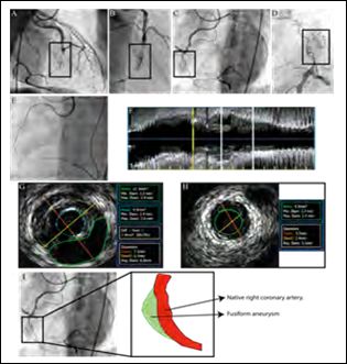
Figure 1: A-D. Evidence of contrast retention in the mid-RCA with a close-up angiog- raphy identifying a contrast retention area (true lumen vs. chronic coronary dissection or aneurysm). E. CART reverse technique was used to approach the chronic total oc- clusion. F-H. IVUS showed a fusiform apparently three-layer wall true aneurysm and lumen diameter 7 mm (orange line). I. Fusiform aneurysm diagram (green) on the native vessel (red).
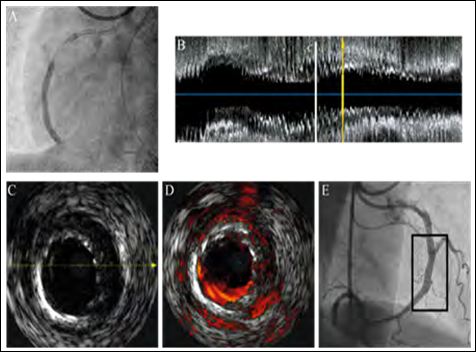
Figure 2: A. CTO was treated successfully with two DES without vascular complica- tion and fusiform aneurysm was apparently isolated. B-D. Final IVUS with successful- ly treatment of the CTO and the fusiform aneurysm with chromaflo confirmation. E. Good angiographic result.
Citation:Salazar C, Salinas P, Gonzalo N, Escaned J (2020) An Unexpected Surprise in the Cath Lab: CTO RCA PCI. J Cardio Cardiovasu Med 4: 011.
Copyright: © 2020 Salazar C, et al. This is an open-access article distributed under the terms of the Creative Commons Attribution License, which permits unrestricted use, distribution, and reproduction in any medium, provided the original author and source are credited.
